
PVR vs. TBI for PAD in Diabetics
PVR vs. TBI: Two Ways to Catch PAD That ABI Misses in Diabetic Patients
Key Takeaway: In diabetic patients with calcified arteries, standard ABI readings can come back falsely normal or falsely elevated. Two exam types solve this problem in different ways. PVR (pulse volume recording, performed with the ABI-Q exam) measures blood volume waveforms and is unaffected by vessel wall stiffness. TBI (toe-brachial index) measures pressure at the toe, where small digital arteries rarely calcify. Both detect PAD that ABI alone would miss. Used together, they confirm findings and close the diagnostic gap.
The Diabetic ABI Problem: When a “Normal” Result Isn’t Normal
Ankle-brachial index has been the standard PAD screening tool for decades. It works by comparing systolic pressure at the ankle to systolic pressure at the arm. In a healthy patient, that comparison is reliable.
Diabetic patients break the model. Chronic hyperglycemia drives calcium deposits into the tunica media of arterial walls, a process known as medial arterial calcification (Monckeberg’s sclerosis). Calcified arteries resist compression. When a blood pressure cuff can’t fully occlude a stiff vessel, the ankle pressure reads artificially high, sometimes above 1.30. A clinician looking at that number might conclude the patient has healthy circulation. Underneath, significant PAD can be present and advancing.
This isn’t a rare edge case. Diabetic patients and patients with chronic kidney disease are exactly the population most likely to have calcified, noncompressible arteries and exactly the population at highest risk for PAD, limb ischemia, and non-healing wounds. Relying on ABI alone in this group leaves a diagnostic blind spot at the worst possible time.
That blind spot is why PVR and TBI exist as complementary exams. Each works around arterial calcification through a different physiological mechanism.
How PVR (ABI-Q Exam) Finds PAD Around the Calcification
Pulse volume recording doesn’t measure pressure against a stiff vessel wall at all. Instead, it uses air-filled cuffs to detect small changes in limb volume that occur with each heartbeat. As blood flows into the limb segment during systole, the limb expands slightly. The cuff senses that expansion and translates it into a waveform.
A healthy limb produces a sharp, well-defined waveform: a fast upstroke, a clear dicrotic notch, and a smooth downstroke. As arterial disease progresses and blood flow becomes more restricted, the waveform flattens and rounds out, losing its notch and its sharp peak. The shape of the wave, not a pressure number against a calcified vessel, is what tells the clinical story.
Because PVR reads volume change rather than occlusion pressure, arterial stiffness from calcification doesn’t distort the result the way it distorts ABI. This is the core reason the ABI-Q exam holds up so well in diabetic patients: it’s a Doppler-free, volume-based assessment that any trained staff member can perform quickly, and it isn’t fooled by a rigid vessel wall.
Strengths of PVR:
- Not affected by arterial calcification, so it stays reliable in exactly the patients where ABI fails
- Fast to perform and doesn’t require a Doppler probe or advanced sonographic skill
- Segmental cuff placement can help localize disease to a general level (thigh, calf, ankle) on multi-level systems
- Well suited to high-volume screening in primary care, podiatry, and wound care settings
Trade-offs of PVR:
- Waveform interpretation is pattern-based, not a hard numeric threshold, so it takes some clinician familiarity to read confidently
- It reflects proximal flow rather than a precise focal lesion location without a full segmental cuff setup
- On its own it doesn’t produce the specific quantitative index that some referral and reimbursement pathways ask for
How TBI Finds PAD Around the Calcification
Toe-brachial index takes a different route to the same goal. Instead of avoiding pressure measurement, it moves the pressure measurement to a location where calcification is far less common. Digital arteries in the toe are smaller and structurally different from the tibial and peroneal arteries at the ankle, and they are much less likely to develop the same medial calcification. That makes toe pressure a more trustworthy number in exactly the patients whose ankle pressures can’t be trusted.
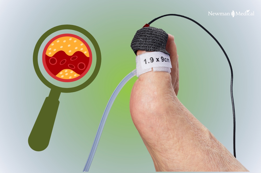
TBI is performed using a small toe cuff paired with a photoplethysmography (PPG) sensor, rather than a Doppler probe. The PPG sensor detects tiny changes in blood volume in the toe tissue as the cuff inflates and deflates, identifying the systolic pressure at which flow returns. That toe pressure is then divided by brachial pressure to produce the toe-brachial index, a number that behaves the way ABI is supposed to: a low index reflects reduced perfusion, and a normal index reflects adequate flow.
Because it produces a quantitative index rather than a waveform shape, TBI is often the exam referenced when calcified arteries make ABI unreliable, and it plays a direct role in wound healing assessment and amputation risk stratification, since toe perfusion correlates closely with the microcirculation needed to heal a distal wound.
Strengths of TBI:
- Toe arteries resist medial calcification, so the index stays clinically meaningful when ABI does not
- Produces a specific, quantitative number rather than a pattern to interpret
- Directly relevant to wound healing potential and surgical planning, since it reflects distal perfusion
- Established diagnostic thresholds support clear documentation and reimbursement
Trade-offs of TBI:
- Takes a bit longer per limb than a quick PVR pass
- Toe cuff placement can be difficult in patients with amputations, deformity, or very small digits
- Cold extremities or vasospasm can transiently affect toe perfusion and skew a single reading
- Requires correctly sized toe cuffs on hand for a range of patients
PVR or TBI? The Honest Answer Is: It Depends on the Question You’re Asking
PVR answers “is blood flow into this limb segment reduced, and where does it change along the leg?” TBI answers “what is the actual perfusion pressure reaching the toe, and is it enough to heal a wound?” Both questions matter in diabetic patients, and they don’t always have the same answer at the same moment in a patient’s disease course.
A patient with an elevated, noncompressible ABI and a flattened PVR waveform already has a strong case for PAD. Adding a TBI on that same patient turns a pattern-based finding into a documented, quantitative index, useful for referral, for surgical planning, and for tracking a wound over time. Running both exams isn’t redundant. It’s how two independent physiological signals confirm the same clinical conclusion, which matters most in the patients whose numbers are hardest to trust in the first place.
Every simpleABI Cuff-Link System Runs Both Exams, Built In
Providers shouldn’t have to choose between purchasing a PVR system and a TBI system, or referring patients out for the exam their equipment doesn’t support. Every simpleABI Automated Cuff-Link System, from the 400CL to the 600CL, comes with the full ABI-Q PVR waveform exam and TBI with PPG built into the same device.
That means a single system can run a fast ABI-Q pulse volume recording as a first pass, add a TBI with PPG when calcification is suspected or confirmed, or run both on the same visit to cross-check findings before a referral. Smart automation handles cuff selection and inflation, full reports support proper reimbursement documentation, and DICOM integration keeps results flowing into the EHR without extra steps. For practices managing diabetic patients, this isn’t a nice-to-have. It’s the difference between catching PAD early and missing it behind a falsely reassuring number.
Request a demo to see ABI-Q PVR and TBI with PPG running on the same Cuff-Link system, or call 800-267-5549 to talk with a Newman Medical representative about which Cuff-Link model fits your practice.