
3-cuff Exam
3-Cuff Segmental Exam
An extension of the ABI exam that adds segmental pressure levels to localize where in the leg an arterial occlusion is occurring — without increasing exam complexity significantly.
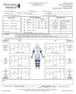 Sample Report
Beyond the ABI: Knowing Where the Problem Is
A standard ABI can confirm PAD is present, but it cannot tell you where the occlusion is. That information matters for treatment planning — whether a patient is a candidate for intervention, and if so, at which level.
The 3-cuff segmental exam adds cuff positions at the thigh and calf to the standard ankle reading, creating pressure gradients that identify the general location of arterial disease: aortoiliac, femoral, or tibial.
How It Works
The protocol is identical to the ABI exam in positioning and patient prep. The Doppler probe remains at the ankle throughout — only the cuff positions change.
Apply 3-Level Cuffs
Cuffs placed bilaterally at the thigh, calf, and ankle. Doppler probe positioned at the ankle (posterior tibial).
Sequential Pressure + PVR
Each cuff level is inflated and deflated in sequence. Pressures and PVR waveforms captured at each of the three levels.
Pressure Gradient Analysis
Software calculates pressure drops between levels. A gradient greater than 20 mmHg between adjacent segments indicates occlusion at that level.
When to Order This Exam
ABI confirms PAD — need to localize the segment
Pre-intervention planning for endovascular or surgical referral
Claudication with ABI in the 0.40–0.90 range
Monitoring disease progression over time
Differentiating aortoiliac from femoral-popliteal disease
Post-intervention follow-up to confirm improvement
Available On
The 3-cuff segmental exam requires a multi-level Cuff-Link system.
Documents
- 3-Cuff Exam Procedure
- Sample 3-Cuff Report — full output example
Training Videos
Questions About This Exam?
Schedule a 30-minute consultation. We'll walk through the protocol, the report output, and which system fits your workflow.